
HBsAgGi
A monoclonal antibody directed against the O-glycosylated PreS2 domain of M-HBs, primarily recognizing infectious HBV particles (DNA virions and RNA virions).
- Product Code: RCA-001
- Package/Specification: 50 μg/tube
- Applicable Genotype: Genotype C
- List Price: USD 700.00-
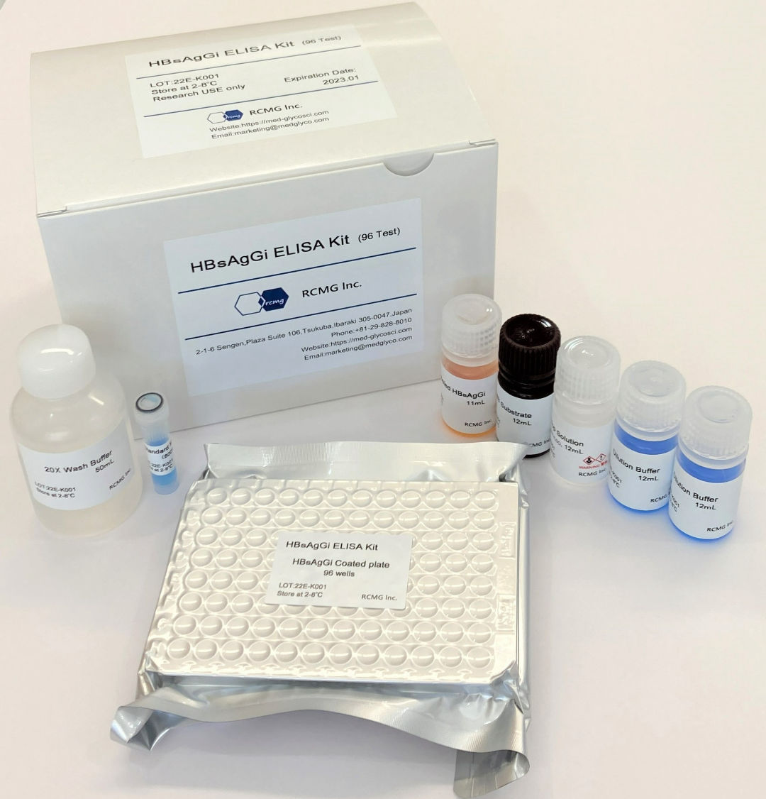
HBsAgGi ELISA KIT
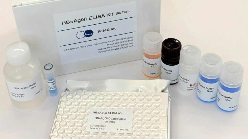
An ELISA kit using a monoclonal antibody directed against the O-glycosylated PreS2 domain of M-HBs, enabling measurement of infectious HBV particles (DNA virions and RNA virions).
- Product Code: RCEK-001
- Package/Specification: 96 Tests
- Applicable Genotype: Genotype C
- List Price: USD 1,000.00-
Features of HBsAgGi
HBsAgGi is an antibody that efficiently recognizes infectious HBV particles of Genotype C, the most prevalent genotype in East Asian countries such as Japan and China.
Unlike conventional HBsAg antibodies, HBsAgGi does not detect non-infectious HBV particles, making it valuable for a wide range of research applications.
- Specimens from chronic hepatitis B patients at various disease stages can be compared against available markers (HBV-DNA, HBV-RNA, qHBsAg, HBcrAg).
- Applicable to a broad range of samples from cell culture supernatants to preclinical study samples. Recognizes the PreS2 domain, enabling detection of HBV strains that evade conventional antibodies due to S domain mutations.
References
| No. | References | Summary |
|---|---|---|
| 1 | Yao B, et al. Antiviral Res. Jan 7, 2025 | HBsAgGi was reported to significantly correlate with HBV RNA in patients with undetectable HBV DNA following nucleos(t)ide analogue therapy, suggesting that HBsAgGi serves as a marker of infectious particles containing HBV RNA in DNA-negative patients. |
| 2 | Okumura T et al, Hepatol Res. Aug 20, 2024. | The potential of HBsAgGi as a novel biomarker was highlighted in an Editorial. |
| 3 | Yuji I, et al. Hepatol Res. May 28, 2024 | A study of 232 HBV-infected patients reported that HBsAgGi levels are a predictive factor for HBsAg seroclearance. |
| 4 | Ivana L et al. Pathogens 2024 Jan 3;13(1):46 | A review article discussed the potential of HBsAgGi as a surrogate marker for HBV RNA and cccDNA. |
| 5 | Kozuka R, al. Hepatol Res. Feb 7, 2024. | A longitudinal analysis of 112 HBV-infected patients reported that the ratio of HBsAgGi to qHBsAg is a predictive factor for hepatocellular carcinoma development. |
| 6 | Okumura T, et al. J Viral Hepat. 30: 731-739, 2023 | A cross-sectional analysis of HBsAgGi in 124 outpatient HBV-infected individuals, with partial longitudinal data, was the first study to suggest an association with cancer onset. |
| 7 | Murata A, et al. BMC Gastroenterol. 22: 270, 2022. | An epoch-making paper describing the development of an ELISA using a monoclonal antibody against HBsAgGi (the “HBsAgGi ELISA Kit” as a research reagent) and the world’s first measurement of serum HBsAgGi levels before and after nucleos(t)ide analogue treatment in 47 treatment-naïve chronic hepatitis B patients. Capture of RNA particles was also reported in this study. |
| 8 | Angata K, et al. Biochim Biophys Acta Gen Subj. 1866:130020, 2022. | The development, characterization, and activity of the monoclonal antibody against HBsAgGi were reported. |
| 9 | Wagatsuma T, et al. Anal Chem. 90:10196-10203. 2018. | The glycan structure preferentially expressed on infectious HBV particles — the O-glycosylation of the PreS2 region of M-HBs (= HBsAgGi) — was identified and reported. |
HBsAgGi ELISA KIT:
An ELISA kit using a monoclonal antibody directed against the O-glycosylated PreS2 domain of M-HBs, enabling measurement of infectious HBV particles (DNA virions and RNA virions).
Product Code: RCEK-001
Package/Specification: 96 Tests
Applicable Genotype: Genotype C
List Price: USD 1,000.00-
ELISA Kit Components
HBsAgGi antibody coated plate:96 well (8-well strip x 12)
Standard M-HBsAg
Dilution Buffer
HRP-labeled HBsAgGi antibody
20X Wash Buffer
TMB Substrate
Stop Solution
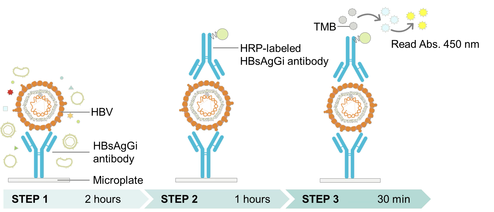
Three-Step Measurement Procedure
- STEP 1: Capture of infectious HBV particles by HBsAgGi antibody
- STEP 2: After washing, reaction with HRP-labeled HBsAgGi antibody
- STEP 3: Addition of TMB substrate → stop reaction → measure absorbance at 450 nm
Safety data sheet (SDS) / User guide
HBsAgGi:
A monoclonal antibody directed against the O-glycosylated PreS2 domain of M-HBs, primarily recognizing infectious HBV particles (DNA virions and RNA virions).
Product Code: RCA-001
Package/Specification: 50 μg/tube
Applicable Genotype: Genotype C
List Price: USD 700.00-
Specifications
⚫︎ Isotype: Mouse IgG1
⚫︎ Light chain: κ (kappa)
⚫︎ Amount: 50 μg
⚫︎ Storage: 4°C (short-term); −20°C (long-term; avoid freeze-thaw cycles)
Applications
Western blotting
Immunoprecipitation & isolation
ELISA
HBV infection inhibition assay
Immunohistochemistry
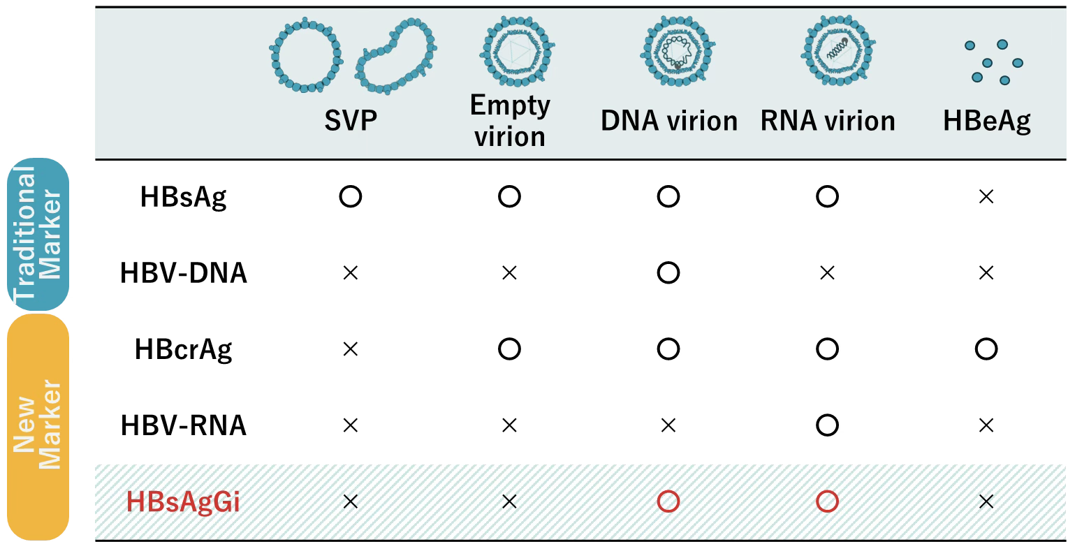
HBsAgGi and HBV Markers
In addition to conventional markers (qHBsAg and HBV DNA), HBcrAg and HBV RNA have recently come into use for assessing the condition of patients with HBV infection. The HBV particles and molecules targeted by each marker including HBsAgGi are shown in the table below.
The HBV particles and molecules targeted by each marker are shown in the table.
○ indicates the target particle/molecule;
× indicates that the marker cannot or is not recommended to recognize that target.
HBsAgGi can detect HBV infectious particles that contain M-HBsAg, such as HBV particles carrying DNA or RNA.